
home > research > Shaped Pulses in EPR
Pulsed EPR method developments
In EPR a magnetic moment of an electron is studied. The magnetic moment is a macroscopic evidence of the quantum mechanics nature of an electron having an intrinsic magnetic moment called spin. One of the simplest way to investigate this spins is turning the spin within a magnetic field. Turning the spin is irradiating a sample containing the spin with electro-magnetic waves (in EPR mostly called microwave) which fulfill the resonance condition of the spin in the applied magnetic field. Depending on the duration of this microwave the spin is turning by a certain angle. Such transmitting microwave for a certain time is called a pulse (rectangular pulse of a certain length). See for example Pulsed EPR in Wikipedia for some detailed explanation [1].
What are shaped pulses?
Rectangular pulses have one basic problem, they actually only act in the assumed way (turning the magnetization by a certain angle)
when the electron spins is on resonance [2]. If the spectral width of the spin assemble is very narrow, the assumption is valid for the whole
spin assemble. However in EPR we deal almost always with spin assembles which are broader than the excitation width of a rectangular pulse.
The problem could be solved, when we do not use one rectangular pulse only, but a combination of rectangular pulses with different frequencies,
amplitudes and phases. This combination of rectangular pulses is called a shaped pulse and is used in NMR experiments since several decades.
Unfortunately for EPR spectroscopy the necessary combination of rectangular pulses exceeded the speed of affordable electronic devices to generate shaped pulses,
until about 10 years ago. Nowadays there are devices which are capable to generate arbitrary waveforms (AWG, arbitrary waveform generator). Such an AWG can generate
pulses with less than 1ns (in our case 0.625ns) changes. This is fast compared to the ‘normal’ length of rectangular pulses (several ns to tens of ns) used in EPR.
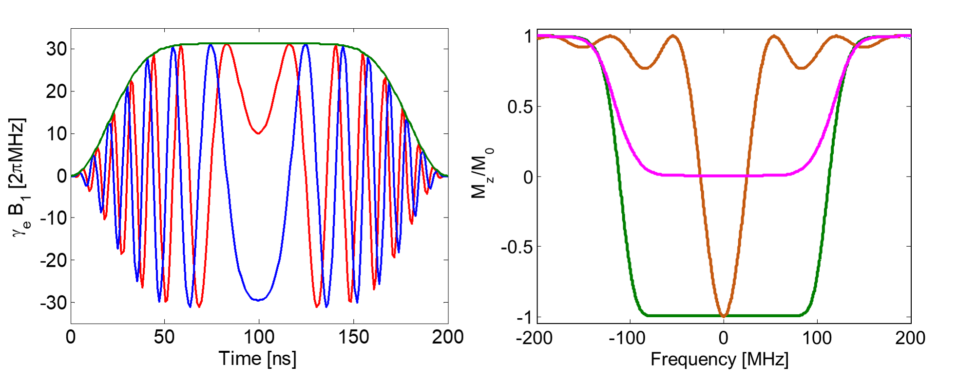
A typical representative of such a class of pulses used in EPR is a WURST pulse (WURST: wideband, uniform rate, smooth truncation). This pulse has a well-defined
excitation bandwidth and can be used as 90° as well as 180° pulse [3].
Projects related to shaped pulses
The pretty new class of shaped pulses gives the opportunity to develop EPR experiments using shaped pulses [4].
Such a development involves understanding the EPR experiment, choosing shaped pulses to enhance the EPR experiment
(compared to the EPR experiment using rectangular pulses) or even using a tool to develop pulse sequences based on optimum control theory.
The application of EPR experiments using shaped pulses to study spin labeled bio-molecules gives for example the opportunity to study,
besides the distance between two spin labels, the orientation as well. Therefore enables to study certain changes of bio-molecules due to
environment or state changes and by this the function of a bio-molecule (e.g. switching of riboswitch, ligand binding of aptamers, different
states of channels or transporters) [5].
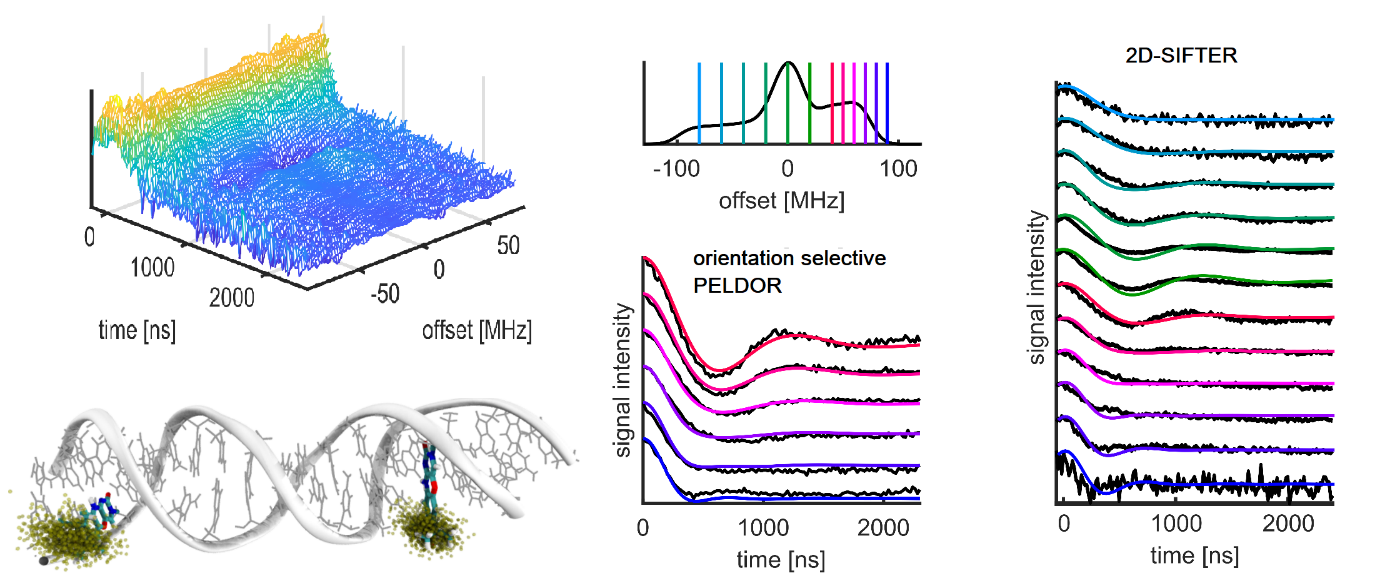
One example of such a experiment is the frequency-correlated 2D SIFTER with broadband pulses at X-band frequencies. At X-band frequencies
the EPR spectrum of nitroxides is governed by the strongly anisotropic nitrogen hyperfine coupling. Broadband shaped pulses allow excitation
of the complete nitroxide EPR spectra. In this case, Fourier transform of the echo signal gives both fast and direct access to the orientation
dependent dipole coupling. Here, we show the application of the frequency-correlated 2D SIFTER experiment with broadband pulses to a double stranded
DNA sample.[5c]
PELDOR measurements at room temperature
PELDOR experiments are typically performed at low temperatures (50K). The main reason for that is the fast transversal relaxation time of nitroxide spin labels at
higher temperature. The fast tumbling of the nitroxide moiety on the sub-ns timescale leads to short relaxation times due to the large anisotropic nitrogen hyperfine
coupling (at X-band frequencies). Several approaches have been undertaken to overcome this limitation.
We were able to perform orientation selective PELDOR measurements at room temperature [6], because the used Ç spin label has no internal
degree of rotational freedom when incorporated into dsDNA. In addition, we used nucleosil beads to prevent tumbling of the DNA molecule itself. The obtained
high-quality PELDOR data at room temperature (Figure 7A) show strong oscillations and orientation selectivity.
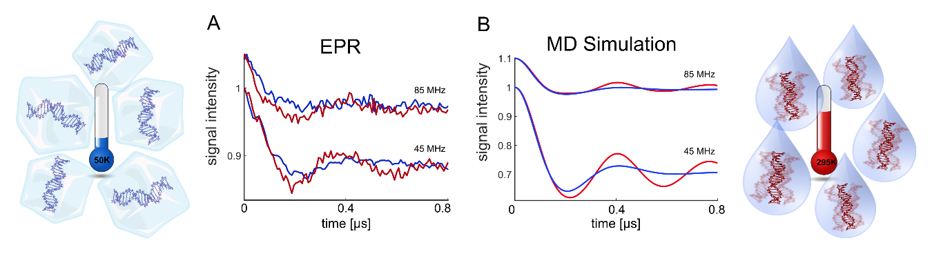
In contrast to all previously performed pulsed dipolar spectroscopy studies at room temperature, our data show a clear difference between the PELDOR time traces measured at low and room temperature. The MD simulations performed by the Hummer group allowed to quantitatively interpret these differences as caused by the fast internal dsDNA dynamics. Whereas the full ensemble of conformers contribute to the PELDOR time traces in frozen solution, only an averaged structure is observed in the PELDOR time traces at room temperature due to the fast time scale of this dynamics. Additionally, we could detect a slight change of the dsDNA structure by the electrostatic interaction with nucleosil at low temperatures. It should be stressed again, that all this information would be impossible to be detected with more flexible spin labels.
References:
[1] wikipedia: Pulsed Electron Paramagnetic Resonance
[2] Spindler, P. E., Schöps, P., Bowen, A. M., Endeward, B. and Prisner, T. F. (2016), Shaped Pulses in EPR. eMagRes., 5, 1477–1492.
doi: 10.1002/9780470034590.emrstm1520
[3] Spindler, P.E., Schöps, P., Kallies, W., Glaser, S. J. and Prisner, T.F. (2017), Perspectives of Shaped Pulses for EPR Spectroscopy. J.Magn.Reson., 280, 30-45.
doi: 10.1016/j.jmr.2017.02.023
[4] Spindler, P. E., Glaser, S. J., Skinner, T. E. and Prisner, T. F.(2013), Broadband Inversion PELDOR Spectroscopy with Partially Adiabatic Shaped Pulses. Angew.Chem.Int.Ed.,52, 3425-3429. doi: 10.1002/anie.201207777
[5] a) Schöps, P., Spindler, P. E., Marko, A., and Prisner, T. F. (2015), Broadband Spin Echoes and Broadband SIFTER in EPR J.Magn.Reson., 250, 55–62. doi:10.1016/j.jmr.2014.10.017
b) Spindler, P.E., Waclawska, I., Endeward, B., Plackmeyer, J., Ziegler, C.M. and Prisner, T. F. (2015), Carr-Purcell Pulsed Electron Double Resonance with Shaped Inversion Pulses. J.Phys.Chem.Lett., 6, 4331–4335.
doi: 10.1021/acs.jpclett.5b01933
c) Bowen, A.M., Erlenbach, N., van Os, P., Stelzl, L.S., Sigurdsson, S.Th. and Prisner, T.F., (2018), Orientation Selective 2D-SIFTER Experiments at X-Band Frequencies. Appl.Magn.Res., 49, 1355-1368.
doi: 10.10.1007/s00723-018-1057-3
[6] Gränz, M., Erlenbach, N., Spindler, P., Gophane, D., Stelzl, L.S., Sigurdsson, S.Th., and Prisner, T.F. (2018), Dynamics of Nucleic Acids at room temperature reveales by pulsed EPR. Angew. Chemie Int. Ed. , 57, 10540-10543
doi: 10.1002/anie.201803682
| Last modified: December 17, 2020 | Impressum / About / Legal Notice | Datenschutzerklärung |
Prisner LOGS
Webmail
Internal
|